Bacteria, singular bacterium, any of a group of microscopic single-celled organisms that live in enormous numbers in almost every environment on Earth, from deep-sea vents to deep below Earth’s surface to the digestive tracts of humans.
Bacteria lack a membrane-bound nucleus and other internal structures and are therefore ranked among the unicellular life-forms called prokaryotes. Prokaryotes are the dominant living creatures on Earth, having been present for perhaps three-quarters of Earth history and having adapted to almost all available ecological habitats. As a group, they display exceedingly diverse metabolic capabilities and can use almost any organic compound, and some inorganic compounds, as a food source. Some bacteria can cause diseases in humans, animals, or plants, but most are harmless and are beneficial ecological agents whose metabolic activities sustain higher life-forms. Other bacteria are symbionts of plants and invertebrates, where they carry out important functions for the host, such as nitrogen fixationand cellulose degradation. Without prokaryotes, soil would not be fertile, and dead organic material would decay much more slowly. Some bacteria are widely used in the preparation of foods, chemicals, and antibiotics. Studies of the relationships between different groups of bacteria continue to yield new insights into the origin of life on Earth and the mechanisms of evolution.
Bacteria lack a membrane-bound nucleus and other internal structures and are therefore ranked among the unicellular life-forms called prokaryotes. Prokaryotes are the dominant living creatures on Earth, having been present for perhaps three-quarters of Earth history and having adapted to almost all available ecological habitats. As a group, they display exceedingly diverse metabolic capabilities and can use almost any organic compound, and some inorganic compounds, as a food source. Some bacteria can cause diseases in humans, animals, or plants, but most are harmless and are beneficial ecological agents whose metabolic activities sustain higher life-forms. Other bacteria are symbionts of plants and invertebrates, where they carry out important functions for the host, such as nitrogen fixationand cellulose degradation. Without prokaryotes, soil would not be fertile, and dead organic material would decay much more slowly. Some bacteria are widely used in the preparation of foods, chemicals, and antibiotics. Studies of the relationships between different groups of bacteria continue to yield new insights into the origin of life on Earth and the mechanisms of evolution.
Bacteria as prokaryotes
All living organisms on Earth are made up of one of two basic types of cells: eukaryotic cells, in which the genetic material is enclosed within a nuclear membrane, or prokaryotic cells, in which the genetic material is not separated from the rest of the cell. Traditionally, all prokaryotic cells were called bacteria and were classified in the prokaryotic kingdom Monera. However, their classification as Monera, equivalent in taxonomy to the other kingdoms—Plantae, Animalia, Fungi, and Protista—understated the remarkable genetic and metabolic diversity exhibited by prokaryotic cells relative to eukaryotic cells. In the late 1970s American microbiologist Carl Woese pioneered a major change in classification by placing all organisms into three domains—Eukarya, Bacteria (originally called Eubacteria), and Archaea(originally called Archaebacteria)—to reflect the three ancient lines of evolution. The prokaryotic organisms that were formerly known as bacteria were then divided into two of these domains, Bacteria and Archaea. Bacteria and Archaea are superficially similar; for example, they do not have intracellular organelles, and they have circular DNA. However, they are fundamentally distinct, and their separation is based on the genetic evidence for their ancient and separate evolutionary lineages, as well as fundamental differences in their chemistry and physiology. Members of these two prokaryotic domains are as different from one another as they are from eukaryotic cells.
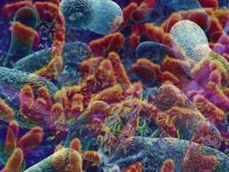
Individual bacteria can assume one of three basic shapes: spherical (coccus), rodlike (bacillus), or curved (vibrio, spirillum, or spirochete). Considerable variation is seen in the actual shapes of bacteria, and cells can be stretched or compressed in one dimension. Bacteria that do not separate from one another after cell division form characteristic clusters that are helpful in their identification. For example, some cocci are found mainly in pairs, including Streptococcus pneumoniae, a pneumococcus that causes bacterial lobar pneumonia, and Neisseria gonorrhoeae, a gonococcus that causes the sexually transmitted disease gonorrhea. Most streptococci resemble a long strand of beads, whereas thestaphylococci form random clumps (the name “staphylococci” is derived from the Greek word staphyle, meaning “cluster of grapes”). In addition, some coccal bacteria occur as square or cubical packets. The rod-shaped bacilli usually occur singly, but some strains form long chains, such as rods of the corynebacteria, normal inhabitants of the mouth that are frequently attached to one another at random angles. Some bacilli have pointed ends, whereas others have squared ends, and some rods are bent into a comma shape. These bent rods are often called vibrios and include Vibrio cholerae, which causes cholera. Other shapes of bacteria include the spirilla, which are bent and rebent, and the spirochetes, which form a helix similar to a corkscrew, in which the cell body is wrapped around a central fibre called the axial filament.
Morphological features of bacteria
THE GRAM STAIN. Bacteria are so small that their presence was only first recognized in 1677, when the Dutch naturalist Antonie van Leeuwenhoek saw microscopic organisms in a variety of substances with the aid of primitive microscopes (more similar in design to modern magnifying glasses than modern microscopes), some of which were capable of more than 200-fold magnification. Now bacteria are usually examined under light microscopes capable of more than 1,000-fold magnification; however, details of their internal structure can be observed only with the aid of much more powerful transmission electron microscopes. Unless special phase-contrast microscopes are used, bacteria have to be stained with a coloured dye so that they will stand out from their background.
One of the most useful staining reactions for bacteria is called the Gram stain, developed in 1884 by the Danish physician Hans Christian Gram. Bacteria in suspension are fixed to a glass slide by brief heating and then exposed to two dyes that combine to form a large blue dye complex within each cell. When the slide is flushed with an alcohol solution, gram-positive bacteria retain the blue colour and gram-negative bacteria lose the blue colour. The slide is then stained with a weaker pink dye that causes the gram-negative bacteria to become pink, whereas the gram-positive bacteria remain blue. The Gram stain reacts to differences in the structure of the bacterial cell surface, differences that are apparent when the cells are viewed under an electron microscope.
Lying outside of this membrane is a rigid wall that determines the shape of the bacterial cell. The wall is made of a huge molecule called peptidoglycan (or murein). In gram-positive bacteria the peptidoglycan forms a thick meshlike layer that retains the blue dye of the Gram stain by trapping it in the cell. In contrast, in gram-negative bacteria the peptidoglycan layer is very thin (only one or two molecules deep), and the blue dye is easily washed out of the cell.
Not only are bacteria able to swim or glide toward more favourable environments, but they also have appendages that allow them to adhere to surfaces and keep from being washed away by flowing fluids. Some bacteria, such as E. coli and Neisseria gonorrhoeae, produce straight, rigid, spikelike projections called fimbriae (Latin for “threads” or “fibres”) or pili (Latin for “hairs”), which extend from the surface of the bacterium and attach to specific sugars on other cells—for these strains, intestinal or urinary-tractepithelial cells, respectively. Fimbriae are present only in gram-negative bacteria. Certain pili (called sex pili) are used to allow one bacterium to recognize and adhere to another in a process of sexual mating called conjugation (see below Bacterial reproduction). Many aquatic bacteria produce an acidic mucopolysaccharide holdfast, which allows them to adhere tightly to rocks or other surfaces.
All living organisms on Earth are made up of one of two basic types of cells: eukaryotic cells, in which the genetic material is enclosed within a nuclear membrane, or prokaryotic cells, in which the genetic material is not separated from the rest of the cell. Traditionally, all prokaryotic cells were called bacteria and were classified in the prokaryotic kingdom Monera. However, their classification as Monera, equivalent in taxonomy to the other kingdoms—Plantae, Animalia, Fungi, and Protista—understated the remarkable genetic and metabolic diversity exhibited by prokaryotic cells relative to eukaryotic cells. In the late 1970s American microbiologist Carl Woese pioneered a major change in classification by placing all organisms into three domains—Eukarya, Bacteria (originally called Eubacteria), and Archaea(originally called Archaebacteria)—to reflect the three ancient lines of evolution. The prokaryotic organisms that were formerly known as bacteria were then divided into two of these domains, Bacteria and Archaea. Bacteria and Archaea are superficially similar; for example, they do not have intracellular organelles, and they have circular DNA. However, they are fundamentally distinct, and their separation is based on the genetic evidence for their ancient and separate evolutionary lineages, as well as fundamental differences in their chemistry and physiology. Members of these two prokaryotic domains are as different from one another as they are from eukaryotic cells.
Individual bacteria can assume one of three basic shapes: spherical (coccus), rodlike (bacillus), or curved (vibrio, spirillum, or spirochete). Considerable variation is seen in the actual shapes of bacteria, and cells can be stretched or compressed in one dimension. Bacteria that do not separate from one another after cell division form characteristic clusters that are helpful in their identification. For example, some cocci are found mainly in pairs, including Streptococcus pneumoniae, a pneumococcus that causes bacterial lobar pneumonia, and Neisseria gonorrhoeae, a gonococcus that causes the sexually transmitted disease gonorrhea. Most streptococci resemble a long strand of beads, whereas thestaphylococci form random clumps (the name “staphylococci” is derived from the Greek word staphyle, meaning “cluster of grapes”). In addition, some coccal bacteria occur as square or cubical packets. The rod-shaped bacilli usually occur singly, but some strains form long chains, such as rods of the corynebacteria, normal inhabitants of the mouth that are frequently attached to one another at random angles. Some bacilli have pointed ends, whereas others have squared ends, and some rods are bent into a comma shape. These bent rods are often called vibrios and include Vibrio cholerae, which causes cholera. Other shapes of bacteria include the spirilla, which are bent and rebent, and the spirochetes, which form a helix similar to a corkscrew, in which the cell body is wrapped around a central fibre called the axial filament.
Morphological features of bacteria
THE GRAM STAIN. Bacteria are so small that their presence was only first recognized in 1677, when the Dutch naturalist Antonie van Leeuwenhoek saw microscopic organisms in a variety of substances with the aid of primitive microscopes (more similar in design to modern magnifying glasses than modern microscopes), some of which were capable of more than 200-fold magnification. Now bacteria are usually examined under light microscopes capable of more than 1,000-fold magnification; however, details of their internal structure can be observed only with the aid of much more powerful transmission electron microscopes. Unless special phase-contrast microscopes are used, bacteria have to be stained with a coloured dye so that they will stand out from their background.
One of the most useful staining reactions for bacteria is called the Gram stain, developed in 1884 by the Danish physician Hans Christian Gram. Bacteria in suspension are fixed to a glass slide by brief heating and then exposed to two dyes that combine to form a large blue dye complex within each cell. When the slide is flushed with an alcohol solution, gram-positive bacteria retain the blue colour and gram-negative bacteria lose the blue colour. The slide is then stained with a weaker pink dye that causes the gram-negative bacteria to become pink, whereas the gram-positive bacteria remain blue. The Gram stain reacts to differences in the structure of the bacterial cell surface, differences that are apparent when the cells are viewed under an electron microscope.
- THE CELL ENVELOPE
Lying outside of this membrane is a rigid wall that determines the shape of the bacterial cell. The wall is made of a huge molecule called peptidoglycan (or murein). In gram-positive bacteria the peptidoglycan forms a thick meshlike layer that retains the blue dye of the Gram stain by trapping it in the cell. In contrast, in gram-negative bacteria the peptidoglycan layer is very thin (only one or two molecules deep), and the blue dye is easily washed out of the cell.
- CAPSULES AND SLIME LAYERS
- FLAGELLA, FIMBRIAE, AND PILI
Not only are bacteria able to swim or glide toward more favourable environments, but they also have appendages that allow them to adhere to surfaces and keep from being washed away by flowing fluids. Some bacteria, such as E. coli and Neisseria gonorrhoeae, produce straight, rigid, spikelike projections called fimbriae (Latin for “threads” or “fibres”) or pili (Latin for “hairs”), which extend from the surface of the bacterium and attach to specific sugars on other cells—for these strains, intestinal or urinary-tractepithelial cells, respectively. Fimbriae are present only in gram-negative bacteria. Certain pili (called sex pili) are used to allow one bacterium to recognize and adhere to another in a process of sexual mating called conjugation (see below Bacterial reproduction). Many aquatic bacteria produce an acidic mucopolysaccharide holdfast, which allows them to adhere tightly to rocks or other surfaces.
- THE CYTOPLASM
- GENETIC CONTENT
- CYTOPLASMIC STRUCTURES
Credits to the owner: http://www.britannica.com/EBchecked/topic/48203/bacteria
